Dental x-ray
Digital technology for minimum exposure to radiation
Dental CT – CBCT – 3D scan of the jaw
Dental CT (CBCT) allows the most detailed three-dimensional insight into the anatomical structures of the jaw and plays a key role in diagnostics, as well as in implantology and orthodontic planning. Unlike Panoramic, which is two-dimensional, Dental CT shows the bone in all three dimensions, as well as its density. In this way, we can detect pathological processes that can remain hidden on 2D images, as well as plan the position, depth and inclination of dental implants in detail. 3D imaging is used in Orthodontic planning as well, especially in the crowded cases, where we decide between expanding the dental arch and teeth extraction.
ŠkodaDENT is equipped with a modern Digital CBCT device that emits the smallest dose of radiation. The device can simultaneously produce 2D and 3D scans, so there is no need for additional scanning. Therefore, with a CT scan of both jaws we offer a FREE Panoramic to our patients.
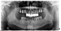
PANORAMIC RADIOGRAPH
Digital Pantomograph, Orthopantomograph or Panoramic radiograph is a panoramic dental X-ray showing the teeth of both the upper and lower jaw simultaneously, along with the surrounding tissues and structures. It is considered to be one of the basic diagnostic tools in dentistry and is often used as an orientation at the first dental examination. It is mandatory when planning the prosthetic therapy, in orthodontics and in oral surgery when planning the dental implant placement and positioning or the extraction of impacted teeth. When it comes to children, panoramic radiographs are safe for them, too, since the radiation dose gets adjusted, i.e. lowered. The only group that should avoid X-rays are pregnant women, especially in the first trimester. Here in Škodadent, we use the latest digital technology, so our X-ray devices use the least possible radiation dose which is not considered harmful.
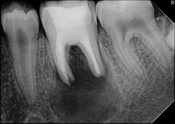
RETROALVEOLAR RADIOGRAPHY
Retroalveolar radiograph, the so-called “small image of the tooth”, enables a detailed view of a couple of teeth and the surrounding bone. It is used when it’s necessary to see a detailed high-quality image of a certain tooth. This kind of radiograph shows the entire tooth from crown to root, and the bone surrounding the tip of the root. A detailed view of the tip of the tooth and the surrounding area is important when planning and controlling the endodontic treatment, because that is the very place where the inflammatory reaction occurs.
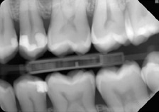
BITE-WING RADIOGRAPHY
This type of radiograph is taken with a patient biting down on a little tab of paper or plastic situated in the centre of the X-ray film, and it shows the crowns of a few posterior and anterior teeth of a certain area in the mouth. Bite-wing radiographs don’t show the area surrounding the tip of the root, but provide a good view of any changes on the teeth crowns. That is why this technique is most commonly used to detect interproximal cavities. In its early stage, this kind of decay can be hard to detect by mere physical examination, but it can be noticed in a bite-wing radiograph. They are recommended to be taken once a year as a part of the annual checkup.